
AI news ·
UToledo researchers develop AI tool that detects osteoporosis risk using existing CT scans
A University of Toledo AI model predicts osteoporosis risk from existing CT scans with about 88% accuracy. Hip fractures alone cost the U.S. $1 billion a year, and the tool aims to catch the disease before a break occurs.
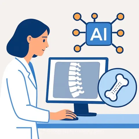
Researchers at the University of Toledo have developed an AI model that analyzes existing CT scans to predict osteoporosis risk, potentially catching the disease years before a fracture occurs. The tool extracts 18 bone-density features from scans patients already have on file and delivers a risk assessment within minutes, offering an alternative to dedicated DEXA scans that often involve scheduling delays.
The team, led by Dr. A. Champa Jayasuriya, professor of orthopaedic surgery and bioengineering, built the model to isolate bone tissue while discarding soft tissue and background imagery. It then combines those extracted features with clinical data points - age, body mass index, and lab markers - to classify risk as high, medium, or low. In testing, the model has shown approximately 88% accuracy, with a goal of reaching 90% in the next development phase.
The cost of silent disease
Osteoporosis often goes undetected until a bone breaks. "Once they have a fracture, then it is very severe, especially a hip fracture," Jayasuriya said. "Sometimes they must get a full hip transplant, at which point they cannot go to work and somebody needs to take care of them. The burden is very high for the patient as well as society - it costs a billion dollars per year in the United States for these hip fractures."
The current standard, a dual-energy X-ray absorptiometry (DEXA) scan, measures bone density but requires a separate appointment. Many at-risk patients never get one until an emergency department visit forces the issue. The UToledo tool is designed to work from imaging data already sitting in hospital systems.
From prototype to clinical testing
The project recently received a $50,000 National Science Foundation grant through UToledo's I-Corps program, which focuses on commercializing academic research. The team will use the funding for further testing and customer discovery with clinicians, radiologists, and potential end users. They have filed a patent application through the university's Technology Transfer Office and are finalizing a manuscript for journal submission.
Shyama Tripathy, an incoming doctoral candidate who worked on the model, pointed to the financial logic behind the approach. "My motivation is that healthcare is really expensive in the U.S.," he said. "By offering this as a service to both physicians and the direct patient, they can save money on expensive exploratory tests."
The team is now working to expand the model beyond upper femur scans to include the hip, spine, and other skeletal areas. They are also addressing a practical hurdle: standardizing performance across CT scanners from different health systems, a necessary step toward broader clinical adoption and eventual FDA review. Norman Rapino, executive director of Rocket Innovations at UToledo, said the I-Corps training "is not only to push stuff out, but to train the next generation of scientists and engineers to do things that create value for society."
Why this matters for IT and development professionals
This project is a concrete example of how AI for healthcare is being built on existing infrastructure rather than requiring new hardware investments. The model works with CT scans that hospitals already generate, extracting structured data from images that were originally captured for other diagnostic purposes. For developers working in medical imaging or clinical decision support, the technical approach - isolating specific tissue types, discarding irrelevant visual data, and combining imaging features with structured clinical variables - mirrors patterns that apply across radiology AI projects. The accuracy targets and standardization challenges also reflect the practical engineering constraints teams face when moving from research prototypes toward FDA-cleared software.