AI matches expert accuracy in hip dysplasia diagnosis-at 30x the speed
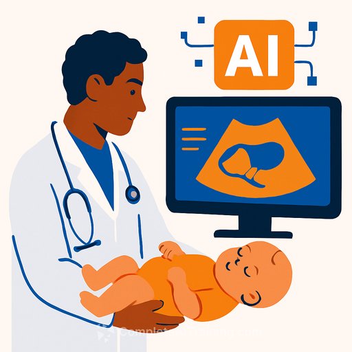
AI systems assessing pediatric hip images are performing on par with clinical experts and doing it about 30 times faster. A review of 23 studies covering more than 15,000 images of children from birth to age 10 found expert-level accuracy with dramatic gains in throughput.
For clinicians, this points to earlier detection, fewer missed cases, and a meaningful cut in time-to-decision-especially in busy services and remote clinics.
Why this matters for care teams
- Earlier intervention: Timely diagnosis can prevent long-term joint problems, reduce chronic pain risk, and avoid major surgery later in life.
- Access at the edge: Nurses, midwives, and junior doctors can capture usable images with minimal training, expanding screening where specialists are scarce.
- Workload relief: Automated triage and measurements mean radiologists focus on complex cases, not volume.
- Fewer appointments for families: Faster, more consistent reads reduce repeat visits and delays.
What the evidence shows
Researchers from the University of Western Australia and the Child and Adolescent Health Service reviewed AI tools that interpret pediatric hip ultrasounds and x-rays. Their synthesis shows accuracy matching clinicians, with dramatic speed gains.
The work was published in the Royal Australasian College of Physicians' Journal of Paediatrics and Child Health. See the journal site for context on current pediatric diagnostics: Journal of Paediatrics and Child Health.
How to pilot this in your service (next 6-12 months)
- Define the first use case: Neonatal hip dysplasia screening and triage for ultrasound and/or x-ray.
- Standards for image capture: Create simple protocols for probe position, angle, and labeling. Build a one-page QC checklist so nurses and midwives can get it right the first time.
- Training at the point of care: Short sessions for junior doctors and midwives on acquiring "AI-ready" images. Emphasize consistent views and minimal artifacts.
- Local validation before deployment: Test on your devices and patient mix. Measure sensitivity, specificity, and time-to-report. Include stress tests with low-quality, reversed, and noisy images.
- Integration into workflow: Connect to PACS/RIS. Route low-confidence or ambiguous cases to radiologists by default. Keep clinicians in the loop for final decisions.
- Safety and governance: Set confidence thresholds, escalation rules, and audit trails. Monitor for automation bias. Establish a review cadence similar to M&M for AI-related cases.
- Equity checks: Track performance by sex, ethnicity, gestational age, and site. Adjust thresholds and retrain if gaps appear.
- Regulatory and privacy: De-identify data, document model versioning, and prepare for device-level approvals where required.
- KPIs that matter: Diagnostic accuracy, time to treatment, reduced repeat imaging, parent visit burden, and cost per case. Report monthly.
- Upskill your team: Give clinicians and imaging staff practical AI literacy to spot failure modes and guide adoption. If helpful, explore role-based AI courses: AI courses by job.
Known limitations and guardrails
- Image quality and angle variance can degrade performance. Standardize acquisition and build QC into intake.
- Generalizability depends on training diversity. Favor models trained on broad, multi-site datasets and keep monitoring for drift.
- Device differences matter. Validate across probes, manufacturers, and software versions.
- Population diversity: Ensure inclusion of preterm infants and varied body types; keep tuning as your data accrues.
- Communication and consent: Be clear with parents about AI-assisted reads and clinician oversight.
What this means for workforce and access
With a projected global shortage of health workers by 2030, shifting image capture and initial triage to trained non-specialists can ease bottlenecks and extend services to rural and low-resource settings. The researchers emphasize this is augmentation, not replacement: AI provides speed and consistency; clinicians provide judgment and accountability.
Adoption outlook
The team expects hospital deployments within five years, pending thorough testing and optimization. Start with controlled pilots, keep a human in the loop, and let real outcomes guide scale-up.
Bottom line
AI for pediatric hip dysplasia can deliver expert-level reads at a fraction of the time. Build simple imaging protocols, validate locally, and deploy with clear guardrails. The payoff: earlier treatment, fewer unnecessary visits, and radiology teams focused where their expertise has the most impact.
Your membership also unlocks:

