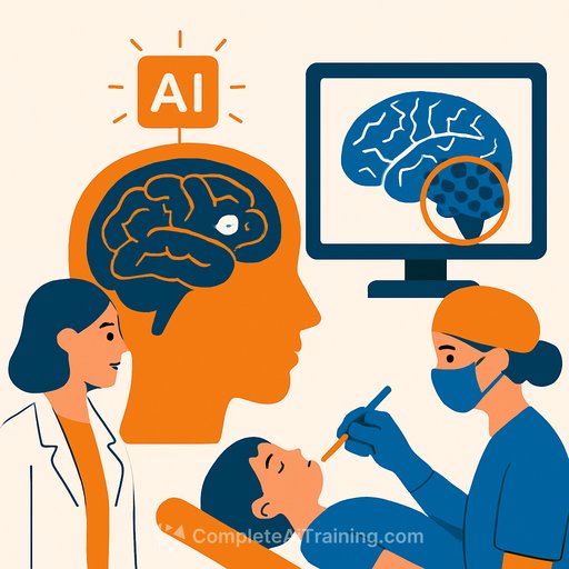
AI tool flags tiny brain malformations in pediatric epilepsy, speeding access to surgery
An artificial intelligence tool developed in Australia can spot tiny, hard-to-detect brain malformations in children with epilepsy. These subtle lesions are often missed on MRI, delaying access to surgery that can dramatically reduce seizures.
This is a practical use case of AI in clinical imaging: sift through large volumes of scan data, highlight suspicious regions, and support a faster, more confident diagnosis. Experts estimate that around three in ten epilepsy cases stem from structural abnormalities in the brain.
Why this matters for care teams
- Earlier detection of subtle lesions: Tiny cortical malformations can be overlooked on initial reads. A second, AI-assisted pass raises the odds of finding them.
- Faster path to surgery: When the lesion is identified, pediatric patients can move more quickly into a surgical workup and, where appropriate, definitive treatment.
- Reduced diagnostic uncertainty: AI acts as an extra set of eyes. It doesn't replace clinicians; it supports them when the imaging is borderline or "MRI-negative."
What this means for your workflow
- Use AI as a decision-support layer: Incorporate it after the initial radiology read to flag regions of interest for review by neuroradiology and the epilepsy surgery team.
- Standardize imaging inputs: High-quality, epilepsy-focused MRI protocols (e.g., high-resolution 3D T1, FLAIR) improve both human and algorithm performance.
- Tighten the referral loop: Positive AI flags should trigger a coordinated review: pediatric neurologist, neuroradiologist, neuropsychologist, and neurosurgeon.
- Document concordance: Track alignment between AI findings, EEG, semiology, and neuropsychology to strengthen surgical candidacy decisions.
Practical adoption checklist
- Clinical governance: Approve AI use under your institution's AI/ML policy with clear responsibility, oversight, and audit trails.
- Validation: Run local retrospective cases to benchmark sensitivity/specificity before clinical rollout; monitor false positives and negatives.
- Integration: Embed into PACS/RIS with structured reporting so AI-marked regions appear seamlessly in the reading workflow.
- Equity and access: Ensure consistent access for pediatric patients across sites; avoid bias by validating across diverse populations and scanners.
- Family communication: Set expectations: AI supports the team; it doesn't give a diagnosis on its own. Share how AI findings fit with the overall pre-surgical evaluation.
- Data protection: Confirm de-identification, secure storage, and compliance with local regulations for any cloud processing.
Clinical caveats
- AI is not a final arbiter: Use it to prioritize review, not to overrule clinical judgment.
- Quality in, quality out: Motion, low-resolution sequences, or inconsistent protocols reduce performance.
- Maintain second reads: Keep human double-reading for challenging pediatric cases, even with AI in place.
Action steps for the next 30 days
- Identify epilepsy-focused MRI protocols used for pediatric cases and standardize across scanners.
- Pilot an AI second-read on recent "MRI-negative" pediatric epilepsy cases and present findings at your MDT.
- Define escalation criteria: when an AI-flag warrants urgent MDT discussion or repeat imaging.
- Set up routine outcome tracking: lesion detection rates, time to surgery, seizure outcomes at 12 months.
Further reading
Build your team's AI capability
If you're tasked with evaluating or implementing clinical AI, upskilling the team shortens the learning curve and reduces deployment risk.
- AI courses by job role for clinicians, analysts, and IT leads
Your membership also unlocks:


