
AI-Based OCT Analysis Pinpoints High-Risk Cardiovascular Patients
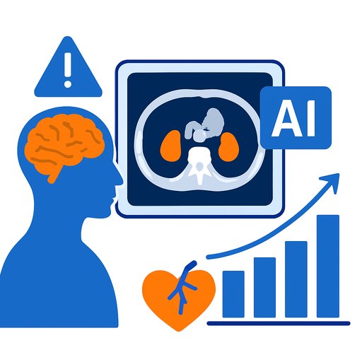
An AI-driven optical coherence tomography (OCT) analysis has demonstrated superior ability in detecting patients at increased risk of adverse cardiovascular events compared to traditional core laboratory OCT assessments. These findings come from the PECTUS-AI study and were shared at the 2025 European Society of Cardiology (ESC) Congress, with results published in the European Heart Journal.
The study highlights the potential of AI to improve patient risk stratification by analyzing OCT images more comprehensively and efficiently than manual methods.
AI Outperforms Core Lab in Identifying Thin-Cap Fibroatheroma (TCFA)
The research team previously developed AI algorithms to automatically interpret OCT data. The PECTUS-AI study took this further by assessing whether AI-identified thin-cap fibroatheromas (TCFAs) correlate with real-world patient outcomes.
They analyzed OCT images from 414 patients who had experienced myocardial infarction, focusing on fractional flow reserve-negative nonculprit lesions. Both an independent core laboratory and the AI-based OCT-AID algorithm evaluated the presence of TCFA.
Key findings include:
- AI detected TCFA in 34.5% of patients, compared to 30% identified by core lab review.
- AI-detected TCFA in the target lesion was significantly linked to adverse outcomes, nearly doubling risk (hazard ratio 1.99; P = .04).
- Core lab TCFA assessment showed no statistically significant association (hazard ratio 1.67; P = .14).
- When the entire vessel pullback was analyzed by AI, the predictive power increased substantially (hazard ratio 5.50; P < .001), with a negative predictive value of 97.6%.
AI Captures the Full Vessel Picture, Not Just the Target Lesion
Manual OCT analysis traditionally focuses on target lesions identified via angiography due to the time-consuming nature of reviewing entire vessels. AI overcomes this by automatically assessing the full vessel pullback, uncovering high-risk plaques that may lie outside the angiographic hotspots.
This broader analysis accounts for the stronger prognostic value observed with AI, as it provides a more complete picture of plaque vulnerability along the vessel.
Clinical Implications and Future Directions
The use of AI in OCT analysis could encourage wider adoption of this imaging modality by simplifying interpretation and reducing variability between observers. Real-time AI tools in catheterization labs may soon enable faster, semiautomatic coronary assessments, personalizing treatment and improving prognostication.
Experts emphasize two main clinical takeaways:
- Automated, reproducible identification of high-risk plaques can standardize patient evaluation in everyday clinical practice.
- Assessing entire vessel segments, rather than isolated lesions, aligns with the systemic nature of coronary plaque vulnerability, potentially guiding preventive interventions.
Such advancements could integrate AI-driven OCT interpretation into routine workflows, bridging imaging data with clinical decision-making.
Funding and Disclosures
The study received partial funding from Abbott Vascular. The researchers involved reported no relevant financial conflicts.
For healthcare and research professionals interested in AI applications in medical imaging and beyond, exploring structured AI education can provide valuable insights and skills. Consider visiting Complete AI Training's latest AI courses for relevant learning opportunities.
Your membership also unlocks: