
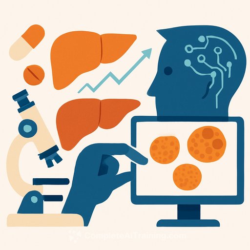
Developing an AI Model for Predicting Drug-Induced Liver Injury Using Liver Organoid Brightfield Images
Drug-induced liver injury (DILI) is a major cause of drug withdrawals and a critical concern in drug safety assessment. Traditional animal models often fall short in predicting human hepatotoxicity due to physiological differences. To address this, researchers have developed an AI-driven approach that leverages brightfield imaging of human liver organoids (HLOs) to predict DILI levels effectively.
Why Use Human Liver Organoids and Brightfield Imaging?
Human liver organoids mimic liver physiology more accurately than animal models or simpler cell cultures. They contain multiple liver cell types and maintain key metabolic functions, including drug metabolism via the cytochrome P450 system. This makes them a closer representation of the human liver environment.
Brightfield imaging offers several advantages over fluorescence microscopy: it is non-destructive, cost-effective, and allows real-time monitoring without additional sample preparation. These features make it ideal for high-throughput screening and continuous observation of organoid morphology.
The AI Model: DILITracer
The AI model, named DILITracer, predicts the level of liver injury—categorized as Most-DILI, Less-DILI, or No-DILI—based on sequential brightfield images of HLOs exposed to drugs. This ternary classification is a first in hepatotoxicity prediction.
DILITracer employs the BEiT-V2 vision transformer, pretrained on 700,000 cell images, to capture 3D morphological features over time. It uses an image-spatial-temporal coding layer to extract and analyze spatiotemporal information from continuous image sequences, linking changes in HLO morphology to DILI severity.
Testing involved 30 compounds from the FDA DILIrank database, covering a range of hepatotoxic potentials and mechanisms. The model achieved an overall accuracy of 82.34%, with a particularly high accuracy of 90.16% in correctly identifying non-hepatotoxic compounds.
Building the DILI Prediction System
- System Construction: HLOs were prepared in a “drug-ready” state and exposed to compounds with known DILI levels. Brightfield images were collected continuously over multiple time points and depths (z-axis). These image sequences were labeled with the respective DILI level for AI training.
- System Application: Unknown compounds were tested on HLOs, and their brightfield image sequences were fed into DILITracer to predict the DILI level.
Comparison with HepG2 Spheroids
To evaluate model performance, two 3D liver models were compared: single-cell-type HepG2 spheroids and multi-cell-type HLOs. Both were exposed to the same 30 compounds, and their morphological changes were analyzed.
The HLO-based model outperformed the HepG2 spheroid model, achieving higher accuracy and better recall, specificity, and precision, especially for the categories of non-DILI and less-DILI compounds. This supports the idea that the physiological complexity of HLOs provides more reliable data for AI-based hepatotoxicity prediction.
Biological Validation
Immunofluorescence confirmed that HLOs contain multiple liver cell types, including hepatocytes, sinusoidal endothelial cells, Kupffer cells, and stellate cells. Importantly, HLOs showed significantly higher expression of key metabolic enzymes (e.g., CYP3A4, CYP1A2) compared to HepG2 spheroids, supporting their suitability for toxicity testing.
Understanding the AI’s Decision Process
Attention heatmaps from the model highlighted that DILITracer focuses on relevant morphological changes in the organoids, such as disintegration or growth inhibition, over time and across image layers. This transparency helps validate that the model bases its predictions on meaningful biological changes.
Practical Implications and Future Use
DILITracer offers a fast, low-cost, and non-invasive method for preclinical hepatotoxicity screening. Its use of clinical data for labeling ensures predictions align closely with human outcomes, reducing reliance on animal testing and improving drug candidate selection.
For IT professionals and developers working in AI-driven biomedical applications, this project exemplifies the integration of advanced computer vision, transformer architectures, and biologically relevant models. It highlights how combining high-content imaging with AI can tackle difficult prediction problems in drug safety.
Those interested in expanding their AI skills in healthcare and image analysis may find relevant courses and training at Complete AI Training.
Summary
- DILITracer uses brightfield images of human liver organoids to predict drug-induced liver injury levels with over 82% accuracy.
- The model leverages a vision transformer pretrained on large cell image datasets to extract spatiotemporal morphological features.
- HLOs outperform HepG2 spheroids as the biological model, providing more clinically relevant toxicity data.
- Brightfield imaging enables non-destructive, real-time, and cost-effective data acquisition suitable for high-throughput screening.
- The model’s interpretability through attention maps aids validation and trust in AI predictions.
This approach represents a practical step toward more accurate and efficient preclinical drug safety evaluation.
Your membership also unlocks: