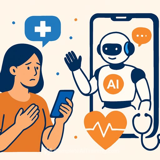
AI in Northeast Ohio Radiology: Faster Reads, Earlier Detection, Human Oversight
Northeast Ohio health systems are using AI to help radiologists read CTs, mammograms, and MRIs faster and with more confidence. The goal is simple: surface what matters quickly, reduce misses, and keep a human expert in control.
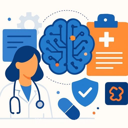
Summa Health, University Hospitals, and Cleveland Clinic are running AI as a second set of eyes. It flags possible abnormalities, prioritizes higher-risk cases, and speeds up image reconstruction so patients spend less time in scanners.
How hospitals are using AI right now
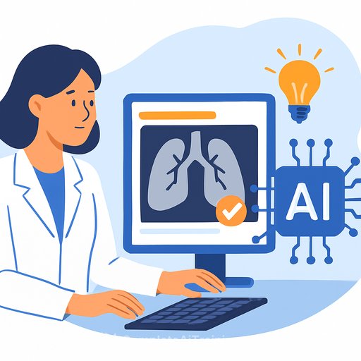
At Summa Health, AI helps detect lung nodules on CT. It highlights suspicious findings for the radiologist, then natural language tools scan reports for keywords like "nodule" and "density" to route higher-risk cases to nurse navigators quickly.
University Hospitals and Summa Health use ClearRead CT (Riverain Technologies), which received FDA clearance in 2016. Studies show it can improve detection of previously missed nodules by 29% and reduce interpretation time by 36%.
Cleveland Clinic uses AI to accelerate MRI image reconstruction across its system (including Akron General, Medina Hospital, and Mercy Hospital). Less time in the bore, fewer motion artifacts, and more patient comfort.
Human oversight stays in the loop
"No AI radiology is running in a way that it is... replacing the radiologist's judgment," said Dr. Leonardo Kayat Bittencourt of University Hospitals. "None of them... runs autonomously in the sense that it... is not adjudicated by a radiologist."
"That AI is never making a diagnosis on its own," said Dr. Po-Hao Chen of Cleveland Clinic. "Every time that it touches a patient's care, it's always been proctored or overseen by the human."
Clinical benefits you can expect
- Earlier lung cancer detection: Many nodules are incidental. In 2025, Summa identified about 3,400 patients with nodules; 357 cancers were found by accident on scans done for other reasons. Catching growth earlier changes outcomes.
- Confidence and speed: AI can review priors while a radiologist reads the current study, outline concerning regions in mammography, and serve as a "sanity check" that nudges certainty when suspicion is already present.
- Less scanner time: Faster MRI reconstruction shortens exams, improves throughput, and reduces patient discomfort.
- Quicker reports: Draft impressions generated by AI cut redundant dictation. Radiologists edit and finalize, preserving clinical judgment.
Workflow gains behind the scenes
- Triage: Algorithms surface high-risk studies first so clinicians act sooner on time-sensitive findings.
- Report mining: NLP tools scan thousands of reports to build actionable lists for navigators and coordinators, reducing manual review time and leakage.
- Standardization: Structured suggestions and consistent highlighting help teams align on thresholds and follow-up protocols.
Risks and failure modes to plan for
- Different error patterns: AI can be wrong in ways humans aren't, and vice versa. Pairing both catches more errors-but only with clear oversight.
- Operational dependencies: If a network hiccup slows an algorithm, reads still proceed, but turnaround may slip if workflows assume instant AI output.
- Validation and drift: Every model needs local vetting, ongoing QA, and performance checks across modalities, vendors, and patient populations.
Patient disclosure: what's being told today
Hospitals generally don't explicitly tell patients that AI assisted in their care behind the scenes. Mammography is a common exception, where departments note that computer-aided detection was used.
The posture across systems is clear: AI augments clinicians. "Nobody's thinking about AI as a means to cut costs or to replace the human expertise," said Bittencourt. The aim is better, faster care with the radiologist firmly in charge.
What this means for your team
- Define the assist: Make it explicit where AI supports detection, triage, reconstruction, and reporting-and where the radiologist must decide.
- Wire the workflow: Build triage queues, navigator handoffs, and EHR flags so findings translate into timely action.
- Stand up QA: Track sensitivity/specificity by indication, site, and scanner; review false positives/negatives in M&M-style huddles.
- Plan for downtime: Create read-without-AI protocols and service-level expectations for delayed results.
- Close the loop: Automate follow-up reminders for nodules and incidentalomas to reduce leakage.
- Educate staff: Train radiologists, technologists, and navigators on indications, limitations, and escalation paths.
KPIs worth tracking
- Time from image acquisition to preliminary read and final report
- Incidental finding capture rate and follow-up completion
- Stage at diagnosis for cancers detected on AI-assisted reads
- Reconstruction time per MRI sequence and scanner throughput
- Change in recall rates and PPV in mammography
- Radiologist and navigator time saved per case
Compliance and evidence
Confirm FDA clearance for each deployed algorithm and keep a local evidence file with performance data and version history. A central registry of AI tools used in your enterprise simplifies audits and quality reviews.
For context on cleared devices and regulatory posture, see the FDA's overview of AI/ML-enabled medical devices here and ACR's AI Central catalog of FDA-cleared radiology tools here.
Bottom line
AI is acting as a reliable second set of eyes in Northeast Ohio radiology-speeding reads, surfacing risk earlier, and shortening MRI exams-while clinicians keep final say. The win comes from pairing algorithms with smart workflows, clear oversight, and tight follow-up processes.
If you're building capability across service lines, start with high-impact use cases (lung nodules, mammography, MRI reconstruction), set guardrails, and measure everything. For broader context and skills, explore AI for Healthcare.
Your membership also unlocks: