AI-Assisted Mammograms: Early Wins, Real Risks, and What Healthcare Teams Should Do Now
During a routine mammogram, a radiologist saw nothing suspicious in Deirdre Hall's images. An AI system disagreed, circling a small area in the upper left breast. Follow-up ultrasound and biopsy found four tumors hidden in dense, overlapping tissue. "This would have been completely missed without the AI," said Dr. Sean Raj of SimonMed Imaging.
Hall's cancer was caught at Stage 1. No lymph node involvement, and treatment decisions could start early. That's the promise many clinicians are seeing: a second set of eyes that never gets tired, especially helpful in dense breast tissue.
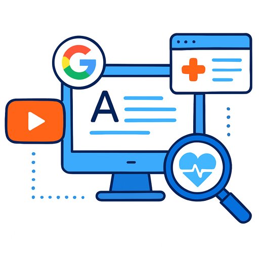
How the technology works
Developers train AI on hundreds of thousands of mammograms. The systems learn patterns that separate benign findings from malignancies. Some tools act like a highlighters, marking areas of concern; others score risk and predict future cancer likelihood.
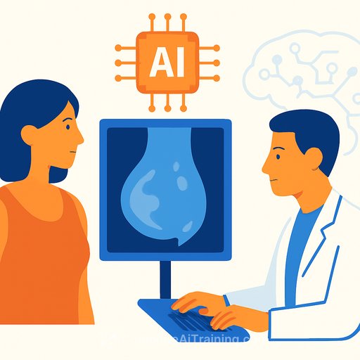
Importantly, these systems do not replace radiologists. FDA rules require a physician to interpret every mammogram. In clinical practice, AI serves as an assistive layer, not an autonomous reader.
What recent studies show
At the University of California, San Francisco, researchers used AI to triage suspicious exams so patients could be seen faster. For people who had breast cancer, average time from screening to biopsy dropped 87% (73 days to 9 days) in a study posted on MedRxiv. It's a preprint, so it still needs peer review, but the time savings are hard to ignore.
Performance varies by product and setting. In a 2024 JAMA Oncology study involving more than 8,800 Swedish women, one system (Lunit) correctly identified cancers 88.6% of the time, with a 7% false positive rate. Another study in Radiology reported AI catching cancers that two radiologists had missed.
Where AI can add value
Dense breasts make screening harder. About 40% of U.S. women fall into this category. As Dr. Otis Brawley of Johns Hopkins put it, finding a small tumor can feel like "trying to find a snowball in a blizzard." AI can help surface subtleties that busy eyes might overlook.
The human-machine combo can also level the field. A RadNet analysis found specialists correctly identified cancers 89% of the time vs. 84% for generalists. With AI, both groups rose to about 93%. As Brawley noted, readings are subjective; AI may reduce disparities in interpretive skill.
Major centers using AI include MD Anderson, Mount Sinai, UPenn's Perelman Center, Siteman Cancer Center, and MedStar Health. The theme is consistent: AI supports the read, a radiologist makes the call.
Limits and concerns you should weigh
Overdiagnosis is real. Some detected cancers would never grow, spread, or threaten life. As Brawley warned, AI could find even more of these indolent tumors, pushing patients into unnecessary procedures and anxiety.
False positives also matter. In the Swedish study, the system flagged about 7% of non-cancers. Across mammography in general, false positives hover around 10%. Each callback means more testing, cost, and stress.
There's also a dependency risk. If clinicians lean too hard on software, errors can creep in. Breast imaging leaders emphasize AI as a tool, not a crutch. And bias remains a concern: if models are trained mostly on images from white women, accuracy may drop for women of color.
Finally, we lack U.S. data showing mortality reduction from AI-assisted screening. A new $16 million, two-year study across seven centers (led by teams at UCLA and UC Davis) aims to fill that gap.
Cost and access
Most academic centers don't charge patients for AI and can't bill insurers because there's no specific code. Some outpatient networks do offer an optional second AI review for a fee (e.g., $40-$50). Ask beforehand so patients aren't surprised.
Dense breasts: why AI may help
Dense tissue is both a risk factor and a masking factor. Overlapping structures can hide tumors on 2D images. In Hall's case, tissue "camouflaged" the cancer, according to Dr. Raj. AI marked the spot anyway, prompting the ultrasound that led to diagnosis.
Screening guidance to use in practice
- USPSTF: Mammogram every other year starting at age 40. See details: USPSTF Breast Cancer Screening.
- American Cancer Society: Ages 45-54, yearly; 55+ every other year or continue yearly based on preference. ACS guidance: ACS Screening & Early Detection.
Practical tips from ACS experts: try to use the same imaging center each year so radiologists can compare prior studies. Ask whether readers are fellowship-trained in breast imaging or primarily read breast exams. Make sure the center can coordinate follow-up imaging if needed.
Your report should note breast density. If it's dense, discuss whether supplemental screening (ultrasound or MRI) makes sense for risk level and local protocols. If a center uses AI, ask how it's integrated, whether it's a standard part of the read, and if there's an optional paid second pass.
Bottom line for healthcare teams
AI is already helping detect cancers that might slip by, especially in dense tissue and busy clinics. It can tighten workflows and support generalists. But it also raises the odds of callbacks and may increase overdiagnosis.
Use AI as an assist, keep the radiologist in the driver's seat, and give patients clear expectations about benefits, risks, timelines, and any extra fees. The clinical gains look promising. The survival impact needs stronger U.S. evidence.
Want to skill up on practical AI for your team?
If you're building internal literacy around AI use in care settings, explore role-based options here: Complete AI Training - Courses by Job.
Additional reading on dense breasts: ACS: Mammograms for Women with Dense Breasts.
Your membership also unlocks: