
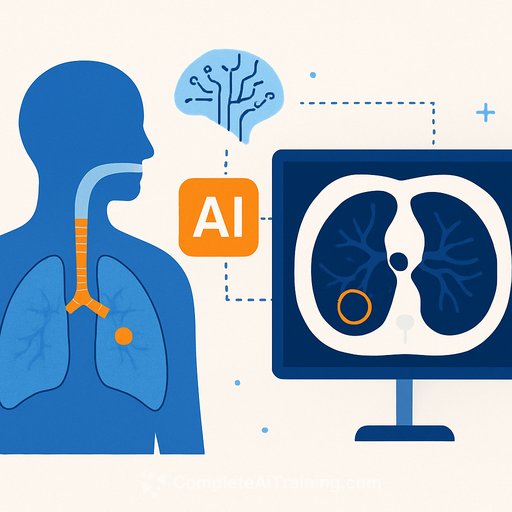
AI flags hard-to-see airway foreign bodies on CT
Researchers at the University of Southampton, working with partners in Wuhan, China, have built an AI model that spots radiolucent airway foreign bodies on CT better than expert readers on case detection. Published in npj Digital Medicine, the system consistently found more true cases than radiologists, offering a reliable second read in high-stakes situations.
"These objects can be extremely subtle and easy to miss, even for experienced clinicians," said PhD researcher Zhe Chen. "Our AI model acts like a second set of eyes, helping radiologists detect these hidden cases earlier and more reliably."
Why this matters for clinical care
Foreign body aspiration (FBA) can present with coughing, choking, and dyspnea-and escalate if missed. Radiolucent material like plant matter or shell fragments often looks faint on CT and is invisible on X-ray, which delays diagnosis.
Up to 75% of adult FBA cases involve radiolucent objects. An assistive tool that lifts case detection can reduce unnecessary morbidity and shorten time to bronchoscopy.
How the model works
The team combined a high-precision airway mapping method (MedpSeg) with a neural network that scans CT volumes for subtle indirect signs of foreign bodies. Training and validation used three independent patient groups totaling more than 400 patients in collaboration with hospitals in China.
The goal: augment human review on difficult reads without changing acquisition protocols.
Study results at a glance
- Test set: 70 CT scans; 14 confirmed radiolucent FBA cases by bronchoscopy.
- Radiologists (3 experts, each >10 years' experience): 36% sensitivity, 100% precision (no false positives).
- AI model: 71% sensitivity, 77% precision (some false positives).
- F1 score: AI 74% vs radiologists 53% (balance of precision and recall).
Bottom line: the AI caught far more true FBA cases while accepting a manageable increase in false positives-useful for triage and second-read workflows.
Clinical takeaways
- Use as a second reader for suspected aspiration with equivocal CT, especially where radiolucent material is likely.
- Expect higher case detection with some false alarms; review AI-flagged regions before recommending bronchoscopy.
- Potential gains: fewer missed FBAs, earlier intervention, and reduced complications.
- This is an assist tool-not a replacement for specialist judgment.
Limitations and next steps
The team plans multi-centre studies with larger, more diverse populations to improve generalisability and reduce bias risk. Local deployment should include prospective audit of sensitivity, precision, and bronchoscopy yield to tune thresholds and workflow fit.
Implementation checklist
- Integrate as an add-on triage/second-read step in PACS or reporting platforms.
- Calibrate thresholds for your patient mix; monitor false-positive rate and downstream bronchoscopy utilization.
- Provide brief reader training on typical AI flags and common pitfalls.
- Track outcomes: time to diagnosis, complication rates, and procedure yield.
For background on airway foreign body aspiration, see an overview on StatPearls (NCBI Bookshelf). Journal context: npj Digital Medicine.
If you're building skills to evaluate or deploy clinical AI, explore practical training: Complete AI Training - courses by job.
Source: University of Southampton
Date: 13.11.2025
Your membership also unlocks: