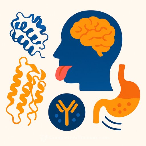
AI clarifies bitter taste receptor structures with AlphaFold3
September 16, 2025
Bitter taste receptors (T2Rs) live far beyond the tongue. They're also expressed in neuropod cells along the gut lining, where they likely influence gut-brain signaling, glucose tolerance, and appetite. A new analysis applies AlphaFold3 (AF3) to map the 3D structures of all 25 human T2Rs and benchmarks those models against AlphaFold2 (AF2) and available cryo-EM structures.
Why this matters
T2Rs are GPCRs that bind a wide range of bitter compounds and couple to α-gustducin. Structural clarity helps explain ligand promiscuity, signaling specificity, and tissue-dependent roles. For researchers, better models mean sharper hypotheses for mutagenesis, ligand screening, and gut-brain axis studies.
What the team did
- Collected amino acid sequences for all human T2Rs from UniProt and predicted structures with AF3.
- Retrieved prior AF2 predictions from the AlphaFold database for head-to-head comparisons.
- Benchmarked against experimentally resolved structures: extensive cryo-EM data for T2R14 (115 structures) and three structures for T2R46 from the Protein Data Bank.
- Used standard toolchains for structure visualization, alignment, and model quality assessment.
Key findings
- AF3 outperformed AF2 consistently across T2Rs, showing tighter agreement with cryo-EM benchmarks for T2R14 and with all resolved structures for T2R46.
- Intracellular regions are more conserved in fold and geometry across the T2R family, aligning with shared G protein coupling demands.
- Extracellular regions vary substantially, consistent with broad ligand recognition and subtype-specific binding pockets.
- Structural clustering split human T2Rs into three groups, offering a practical scaffold for comparative studies and ligand design strategies.
"The expression of bitter taste receptors in the gastrointestinal tract indicates roles in the gut-brain axis, glucose tolerance, and appetite regulation. Structural clarity provides a stronger basis to probe these functions," notes Professor Naomi Osakabe of Shibaura Institute of Technology.
Implications for research and development
- Ligand modeling: Prioritize extracellular pocket mapping and flexible docking across the three structural clusters to triage thousands of bitter compounds.
- Mutagenesis planning: Focus scans on extracellular residues for ligand selectivity and on conserved intracellular motifs for signaling efficiency.
- Gut-brain studies: Use improved models to design probes that selectively engage T2Rs in neuropod cells, then track neural and metabolic readouts.
- Drug discovery: Exploit subtype differences to engineer bitter-mimetic therapeutics or blockers relevant to appetite and glucose control.
- Validation loop: Pair AF3-guided hypotheses with cryo-EM, crosslinking-MS, or deep mutational scanning to confirm binding modes and conformational states.
Data and sources
- Paper: The three-dimensional structure prediction of human bitter taste receptor using the method of AlphaFold3, Current Research in Food Science (2025). DOI: 10.1016/j.crfs.2025.101146
- Experimental structures referenced: Protein Data Bank
Practical next steps
- Build an internal T2R model set using AF3 and annotate extracellular pocket features across the three clusters.
- Run ligand docking panels guided by known bitterant chemotypes; validate predictions with targeted mutagenesis and signaling assays.
- For gut-brain work, test T2R-selective ligands in neuropod cell systems and integrate neural tracing or electrophysiology.
If you're integrating AI into structural biology and screening workflows, explore applied training and certifications: Latest AI courses
Your membership also unlocks:


