Exploring AI-Based Analysis of Histopathological Variability in Salivary Gland Tumours
Salivary gland tumours (SGTs) are a diverse group of neoplasms with overlapping histological features, making their diagnosis challenging. Accurate identification is crucial due to differences in clinical behavior and treatment strategies. Traditional methods often rely on special stains, immunohistochemistry, and molecular tests, which can be costly, time-consuming, and inaccessible in many settings.
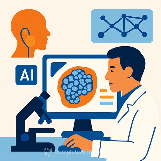
This article presents a study examining the use of artificial intelligence (AI), specifically machine learning (ML) and deep learning (DL) techniques, to differentiate between benign and malignant SGTs, subtype malignant tumours, and grade tumour severity using digitised whole-slide images (WSIs) stained with Haematoxylin and Eosin (H&E).
Study Overview and Methods
The research utilized 320 scanned WSIs encompassing benign tumours like pleomorphic adenoma (PA) and basal cell adenoma (BCA), alongside malignant subtypes such as mucoepidermoid carcinoma (MEC), adenoid cystic carcinoma (AdCC), acinic cell carcinoma (ACC), and carcinoma ex pleomorphic adenoma (Ca-ex-PA). Expert pathologists confirmed diagnoses before slide digitization.
Using QuPath, an open-source image analysis platform, regions of interest (ROIs) were manually selected and analyzed to extract features like cellularity, nuclear and cytoplasmic morphology, and staining optical densities. These features served as inputs for ML classifiers built with Python's Scikit-learn. The study developed three primary classifiers:
- Benign vs. Malignant (BvM) classifier: Differentiates benign from malignant tumours.
- Malignant Subtyping (MST) classifier: Identifies the four most common malignant SGT subtypes.
- Tumour Grading (TG) classifier: Grades tumours as low or high grade for MEC and AdCC subtypes.
For comparison, deep learning models including ResNet-18, ResNet-50, EfficientNet-B0, and EfficientNet-B3 were trained on image patches extracted from the same ROIs.
Key Findings
Classifier Performance: The ML classifiers outperformed DL models across all tasks. The BvM classifier achieved an F1 score of 0.95, surpassing the best DL model’s 0.87. Similarly, the MST classifier reached an F1 score of 0.95, while DL peaked at 0.60. The TG classifier showed an F1 of 0.87 compared to DL’s 0.70.
External validation on independent cohorts confirmed the robustness of the ML classifiers, with F1 scores of 0.87 for both BvM and TG tasks. DL models showed lower generalizability in these settings.
Feature Insights: Several histological features significantly contributed to the classifiers’ decisions, including:
- Nuclear circularity and haematoxylin optical density
- Cytoplasmic eosin optical density
- Nucleus-to-cell area ratio
- Cellularity and spatial distribution of cells
These features differed notably between benign and malignant tumours, among malignant subtypes, and between low- and high-grade tumours.
Spatial Analysis: Quantitative spatial metrics such as centroid distances between cells and Delaunay triangulation revealed distinct patterns. Malignant tumours exhibited higher cellularity and closer intercellular distances compared to benign tumours. Among malignant subtypes, ACC showed the highest number of neighboring cells, while MEC had the largest mean triangle area in spatial clusters.
Implications for Clinical Practice
Applying AI-based analysis to digitized histopathology slides offers an automated, objective method to assist pathologists in diagnosing and grading SGTs. This approach can potentially reduce reliance on ancillary tests, accelerate diagnosis, and improve consistency in settings with limited resources.
While deep learning models are popular, this study highlights that interpretable machine learning methods combined with well-curated feature extraction can achieve superior accuracy and transparency, which is critical for clinical adoption.
Further research involving larger, multicenter datasets is necessary to validate these findings and explore integration into pathology workflows.
Additional Resources
- Complete AI Training – Explore courses on AI applications in healthcare and pathology.
- National Cancer Institute: Diagnosis and Staging – Information on cancer diagnosis methods.
Your membership also unlocks:

