
Beyond amyloid plaques: AI reveals hidden chemical changes across the Alzheimer's brain
Date: March 1, 2026
Source: Rice University
Alzheimer's isn't just a plaque story. A new, dye-free molecular atlas from Rice University maps an Alzheimer's brain (in an animal model) and shows sweeping, uneven chemical shifts that stretch far beyond amyloid deposits.
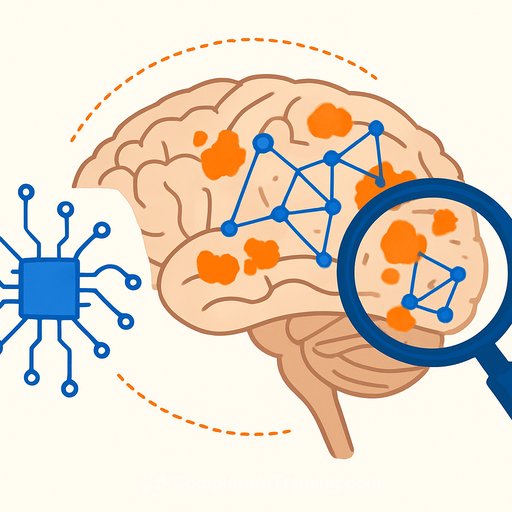
Using hyperspectral Raman imaging paired with machine learning, the team captured a whole-brain view of molecular changes. Key memory regions showed large shifts in cholesterol and energy-related molecules, pointing to a broader metabolic disruption.
What they built
Hyperspectral Raman imaging reads the chemical "fingerprints" of tissue with a scanning laser-no dyes, tags, or labels. Instead of a single spectrum, it collects thousands across entire brain slices to assemble high-resolution chemical maps.
Because the approach is label free, the tissue chemistry is recorded without perturbation. That's useful when the goal is to discover signals you didn't think to look for.
The pattern that stood out
Alzheimer's-linked chemistry was patchy, not uniform. Some regions shifted strongly; others looked closer to healthy tissue. That gradient helps explain why symptoms progress the way they do and why single-target treatments struggle.
The hippocampus and cortex-core memory circuits-showed the biggest differences. Two standouts: cholesterol (cell structure, synapse function) and glycogen (local energy reserve) varied by region, hinting at disrupted structural integrity and energy balance.
The AI stack that made it work
The data volume was massive. Unsupervised models first grouped tissue by molecular signatures-no labels, no assumptions. Supervised models then learned to separate Alzheimer's from control and quantify which regions carried the strongest disease signal.
If you're building similar pipelines, start with unsupervised discovery to avoid bias, then layer supervised models for classification and region-level attribution. For practical training resources, see AI for Science & Research.
Key takeaways for researchers
- Alzheimer's chemistry extends beyond plaques; treat it as a system-level, spatially heterogeneous process.
- Memory hubs (hippocampus, cortex) show pronounced shifts in cholesterol and glycogen-watch lipids and energy metabolism.
- Label-free spectroscopy can surface signals that staining might miss, especially for early or noncanonical changes.
- Region-specific models may outperform whole-brain averages for prediction, staging, and treatment response.
How to apply this in your lab
- Broaden sampling: map multiple regions, not just plaque-rich zones. Include white matter and association cortices.
- Add a label-free modality (e.g., Raman) next to histology, lipidomics, and metabolomics for cross-validation.
- Use unsupervised clustering to reveal spatial motifs, then supervised learning for region-wise disease scores.
- Track cholesterol and glycogen alongside amyloid and tau; align spectra with targeted assays for ground truth.
- Integrate with spatial transcriptomics or proteomics to link metabolic shifts to pathways and cell types.
Important caveats
- Animal model: translational work is required to confirm patterns in human brains and across disease stages.
- Correlation ≠ causation: lipid and glycogen shifts could be drivers, responses, or both.
- Throughput and standardization: hyperspectral datasets are large; pipelines need QC, calibration, and sharable benchmarks.
Where this could lead
Whole-brain chemical maps can support earlier risk flags, region-specific staging, and stratification by metabolic phenotype. They also offer a way to monitor interventions targeting lipids, mitochondria, or glial energy support-not just plaque load.
The work appears in ACS Applied Materials & Interfaces and was supported by the National Science Foundation (2246564, 1934977), the National Institutes of Health (1R01AG077016), and the Welch Foundation (C2144). For clinical context on Alzheimer's biology, see the National Institute on Aging's overview here.
Bottom line
If you study or treat Alzheimer's, widen the frame. Map chemistry across the brain, pair label-free imaging with ML, and follow the metabolism-especially in memory circuits. Plaques matter, but the metabolic terrain may tell you where the disease is headed next.
Your membership also unlocks: