
A UCLA-led research team has built a platform that merges 3D bioprinting, label-free imaging, and AI to track how tumor organoids respond to drugs in real time. The system, announced June 22, 2026, by the UCLA Health Jonsson Comprehensive Cancer Center, processes patient-derived cancer cells into uniform organoid arrays and uses quantitative phase imaging to measure growth without chemical dyes. The result is a high-throughput, single-organoid view of drug sensitivity that could reshape how clinicians select cancer treatments before a patient ever starts a regimen.
How the integrated platform works
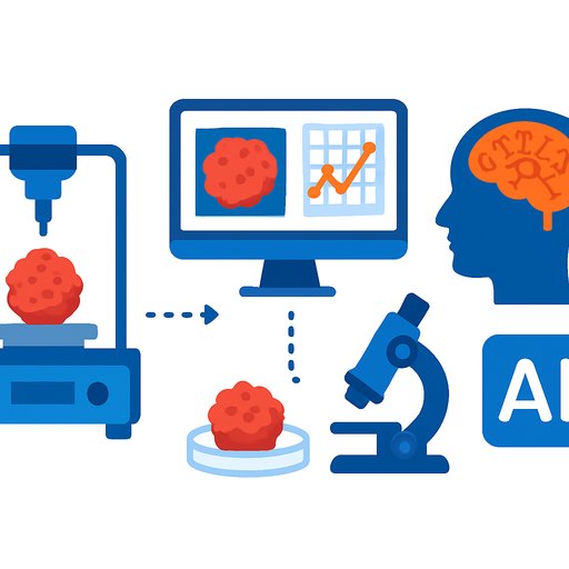
Researchers generate tumor organoids from a patient's own cancer cells and embed them in extracellular matrix constructs. High-speed quantitative phase imaging then captures continuous growth data - tracking biomass and structural dynamics - without fluorescent labels or stains. This non-destructive imaging keeps the samples intact for longer observation windows, which matters when the goal is to catch slow-emerging resistance patterns.
Deep learning-based segmentation and machine learning algorithms process the image streams into structured datasets. The computational pipeline quantifies drug response at the level of individual organoids across thousands of samples. Rather than averaging a bulk measurement across a dish of cells, the system flags which specific organoids stop growing, which continue, and which show partial or delayed responses.
Detecting rare resistant populations
The platform's resolution lets scientists isolate small subpopulations of tumor cells that survive a drug that kills most of the mass. These rare resistant clones often drive relapse, yet they are invisible in conventional viability assays that report a single mean value. By tracking each organoid over time, the system builds a dynamic response profile - not a single endpoint snapshot.
Dr. Michael Teitell, director of the UCLA Health Jonsson Comprehensive Cancer Center and co-senior author of the study, described the shift in logic. "Instead of asking whether a drug works on average for a large number of tumor cells, we can now determine which specific organoids respond and which do not, and, ultimately, have an approach to determine the underlying reasons for unique response profiles," he said.
Validation and scale
The team validated the platform on established cancer cell lines and patient-derived samples. The system screened hundreds of potential therapies simultaneously, showing it can handle the combinatorial load that personalized medicine demands. Contributors from the University of Colorado School of Medicine and Virginia Commonwealth University's Massey Comprehensive Cancer Center collaborated on the work. Funding came from the National Institutes of Health, the National Science Foundation, the Department of Defense, and the Air Force Office of Scientific Research.
Dr. Teitell added that the approach directly targets the gap between population-level trial data and an individual patient's tumor biology. "This allows us to measure drug responses across thousands of individual organoids, detect rare resistant tumor populations, track growth and treatment responses over time, and better predict which therapies may work for a particular patient," he said.
Why this matters for science and research professionals
The platform collapses three workflows - bioprinting, live-cell imaging, and AI-driven analysis - into one continuous loop. For researchers designing drug screens, that means fewer manual transfer steps, less batch-effect noise, and a data structure that captures heterogeneity from the start. The label-free imaging also eliminates dye-related toxicity and photobleaching confounders, which is a persistent problem in long-duration live-cell assays.
For teams working at the intersection of AI for Science & Research and oncology, the study offers a concrete, validated example of how deep learning segmentation can move beyond image classification into quantitative pharmacology. The same computational principles - segment, track, quantify - apply to organoid models in other solid tumors, and the open questions about rare resistant clones are as relevant in pancreatic and ovarian cancer as they are in the melanoma and colorectal models used here. Professionals who build or evaluate similar high-content screening pipelines will recognize the practical win: the system generates single-organoid longitudinal data without adding labor-intensive sample preparation steps that often limit throughput in academic labs.
Your membership also unlocks: