
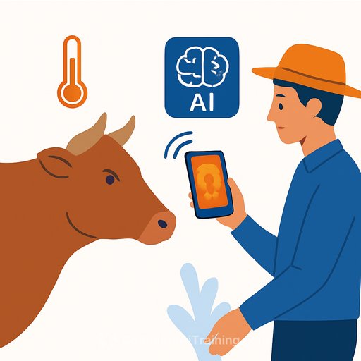
AI estimates a cow's temperature from a single photo
Fever is one of the earliest flags for disease in cattle. The problem: getting accurate readings at scale usually means restraint, contact thermometers, or costly thermal setups. An image-based AI approach changes the workflow by reading body temperature from a standard photo.
This isn't magic. It's a computer vision pipeline trained on images paired with ground-truth temperatures. The model learns visual cues in specific regions of the head-typically the eye area and muzzle-then accounts for ambient conditions to infer core temperature within a practical margin.
How the system works
- Data pairing: Each image is linked to a reference temperature (rectal or calibrated thermal reading). This teaches the model what "fever" looks like in a normal photo.
- Region-of-interest detection: The pipeline locates vascular-rich areas that correlate best with core temperature.
- Context inputs: Ambient temperature, humidity, wind, and time of day can be added as features to improve estimates.
- Calibration: A light calibration step aligns predictions across different camera types and lenses.
How accurate is it?
Trials typically compare model outputs against standard measurements across breeds and environments. Results are often reported as mean absolute error and the share of readings within a clinically useful band (for example, ±0.5°C). Performance can differ by lighting, distance, and whether the animal's face is clean and unobstructed.
Expect better results in controlled lighting, close range, and with consistent camera parameters. Dirty muzzles, occlusions, glare, and extreme ambient conditions tend to degrade estimates.
Why this matters
- Non-contact screening: Reduce stress on animals; no restraint required for a quick check.
- Throughput: Move through large herds faster than manual thermometer checks.
- Cost control: Use commodity cameras or smartphones instead of specialized thermal gear for every station.
- Earlier detection: Flag animals for follow-up before symptoms spread across a pen.
Study design notes researchers care about
- Sampling: Include multiple breeds, ages, and housing conditions; balance indoor and outdoor scenes.
- Ground truth: Use consistent reference methods; log time-lags between imaging and measurement.
- Repeatability: Evaluate day-to-day drift and cross-camera stability.
- Generalization: Test on new farms and unseen lighting to check for overfitting to background cues.
- Uncertainty: Provide per-sample confidence or prediction intervals to guide triage decisions.
Deployment on farm
- Hardware: Mid-range smartphones or IP cameras can work; prioritize stable focus, short shutter times, and consistent distance.
- Workflow: Capture during feeding or milking when heads are forward; standardize angle and range.
- Edge vs. cloud: On-device inference reduces bandwidth; cloud makes fleet updates easier. Many teams run a hybrid.
- Alerts: Trigger a flag when predicted temperature crosses a threshold; route the animal for confirmatory checks.
- Records: Sync to herd-management software for longitudinal trends and biosecurity reports.
Limits to keep in mind
- Extreme heat or cold can skew readings if environmental features are missing or mis-specified.
- Water, mud, or eye discharge can hide cues the model relies on.
- Lens differences and compression can introduce bias without calibration.
- Edge cases (calves vs. mature cattle, certain breeds) may need targeted data to hold accuracy.
Future directions
- Expand datasets across seasons, geographies, and breeds to strengthen external validity.
- Automate camera calibration and self-checks to reduce maintenance.
- Add multitask outputs: respiration rate, cough detection, or behavior anomalies from the same video feed.
- Integrate uncertainty estimates with active learning so the system asks for human validation on atypical samples.
Practical checklist for teams building this
- Define a clinical tolerance (e.g., ±0.5°C) and design your loss/metrics accordingly.
- Collect paired data under varied lighting; log ambient conditions for each capture.
- Use a two-stage model: detector for regions of interest, regressor for temperature.
- Calibrate across devices; include a camera-ID embedding or per-device normalization.
- Report per-subgroup performance; do not rely on a single averaged metric.
- Ship with a confidence score and a simple operator guide for camera distance and angle.
Additional reading
Skills and tools
If you're building image-based diagnostics or farm analytics, a structured learning path helps. See curated options for computer vision, MLOps, and deployment:
Bottom line: a single-photo temperature estimate won't replace confirmatory clinical checks, but it's a practical screen for herd health. Use it to prioritize attention, reduce handling, and capture objective data at scale.
Your membership also unlocks: