
AI news ·
Scientists from India and UK develop AI tool that maps tumours and flags uncertainty in tissue analysis
HISTO-UNet, developed by researchers from India and the UK, detects tumours in tissue images and flags where its confidence is low. No existing diagnostic AI had combined shape accuracy with uncertainty signalling until now.
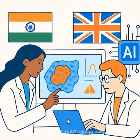
AI Tool Flags Diagnostic Uncertainty in Tissue Analysis
Researchers from institutions in India and the UK have developed an AI system that identifies tumours and glands in microscopic tissue images while signalling where it lacks confidence. The tool, called HISTO-UNet, combines shape preservation with uncertainty quantification-a dual approach absent from existing diagnostic systems.
The team included scientists from the Indian Institute of Science Education and Research Bhopal, Maulana Azad Medical College, Jawaharlal Nehru Cancer Hospital and Research Centre Bhopal, All India Institute of Medical Sciences Bhopal, and the University of Oxford.
How the system works
HISTO-UNet uses a neural network trained with topology-preserving constraints-mathematical rules that force the AI to recognise the skeleton and exact centre of structures rather than producing fragmented shapes or false connections.
The system processes each image 25 times and measures variance across runs to quantify two types of uncertainty. One captures image fuzziness from lab staining techniques; the other measures the AI's own confidence gaps. This dual-layered approach tells doctors exactly where to scrutinise.
Standard algorithms like UNet provide a single prediction with no reliability indicator. Some recent models address shape errors or calculate uncertainty separately, but none had combined both functions until now.
Performance and clinical impact
HISTO-UNet outperformed standard models across three major medical datasets in testing. The trade-off: processing each image multiple times takes longer than simpler systems.
In practice, pathologists reviewing hundreds of tissue samples daily face fatigue-driven errors. By automatically highlighting ambiguous regions, the system converts exhaustive searching into targeted review of difficult cases. This could accelerate diagnosis and reduce oversight mistakes.
Learn more about AI for Healthcare and AI for Science & Research.